45 label thoracic cavity
Solved Correctly label the following anatomical features of | Chegg.com Correctly label the following anatomical features of the thoracic cavity. (Not all words will be used.) Left lung Apex of heart Parietal pleura Fibrous pericardium Superior vena cava Interior vena cava Aorta Pulmonary trunk Base of heart Correctly label the following external anatomy of the anterior heart. (Not all terms will be used.) Body Cavities and Organs | Biology Dictionary The abdominal cavity is where the majority of the body's organs lie. These are sometimes referred to as the "viscera", and they include organs like the liver, stomach, spleen, pancreas, kidneys and others involved in digestion, metabolism, and filtering of the blood.
Body Cavities and Membranes - Anatomy and Physiology Notes The thoracic cavity, also called the chest cavity, sits superior (higher) to the abdominopelvic cavity, and it contains organs such as the heart, lungs, trachea, and esophagus. It can be subdivided into three main portions: The left pleural cavity, which houses the left lung

Label thoracic cavity
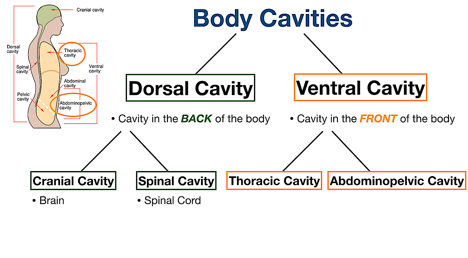
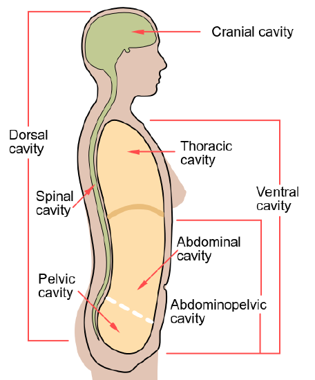
Body Cavities and Membranes: Labeled Diagram, Definitions - EZmed Body Cavities Labeled Diagram: The dorsal cavity is located in the back of the body (red/stars) and houses the central nervous system including the brain and spinal cord. Ventral Cavity The ventral cavity is the cavity located in the front of the body, which makes sense because ventral means front or anterior. Anatomy of the Thoracic Cavity & Diaphragm - YouTube May 16, 2015 ... This video is about anatomy of the thoracic cavity and diaphragm.University of Sulaimani / College of Medicine. Anatomy, Thorax - StatPearls - NCBI Bookshelf - National Center for ... The thoracic cavity contains organs and tissues that function in the respiratory (lungs, bronchi, trachea, pleura), cardiovascular (heart, pericardium, great vessels, lymphatics), nervous (vagus nerve, sympathetic chain, phrenic nerve, recurrent laryngeal nerve), immune (thymus) and digestive (esophagus) systems.
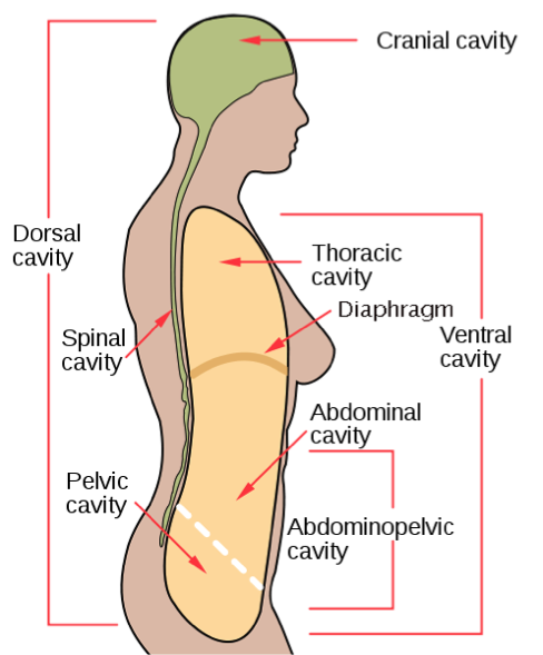
Label thoracic cavity. The Human Body Cavities - Course Hero Cranial cavity-the space occupied by the brain, enclosed by the skull bones. Spinal cavity-the space occupied by the spinal cord enclosed by the vertebrae column making up the backbone. The spinal cavity is continuous with the cranial cavity. Ventral body cavity-the thoracic cavity, the abdominal cavity, and the pelvic cavity in combination. Fetal Pig Dissection - University of Hawaiʻi Figure 9. Mid-line thoracic cut. A cut is made on the side of the animal from the point just posterior to the diaphragm dorsally. A similar cut is made on the other side. These two cuts will enable you to spread open the abdominal cavity. Figure 10. Opening the abdominal cavity. Mouth and Neck Region Thorax: Anatomy, wall, cavity, organs & neurovasculature | Kenhub All the thoracic arteries originate from the aorta and the three largest ones are the brachiocephalic trunk, left common carotid artery, and left subclavian artery. Several visceral arteries also supply various thoracic organs including: bronchial, esophageal, pericardial, and several small mediastinal arteries. 756 Thoracic Cavity Images, Stock Photos & Vectors - Shutterstock Find Thoracic Cavity stock images in HD and millions of other royalty-free ... Thoracic cavity vector illustration drawing labeled diagram Stock Vector ...
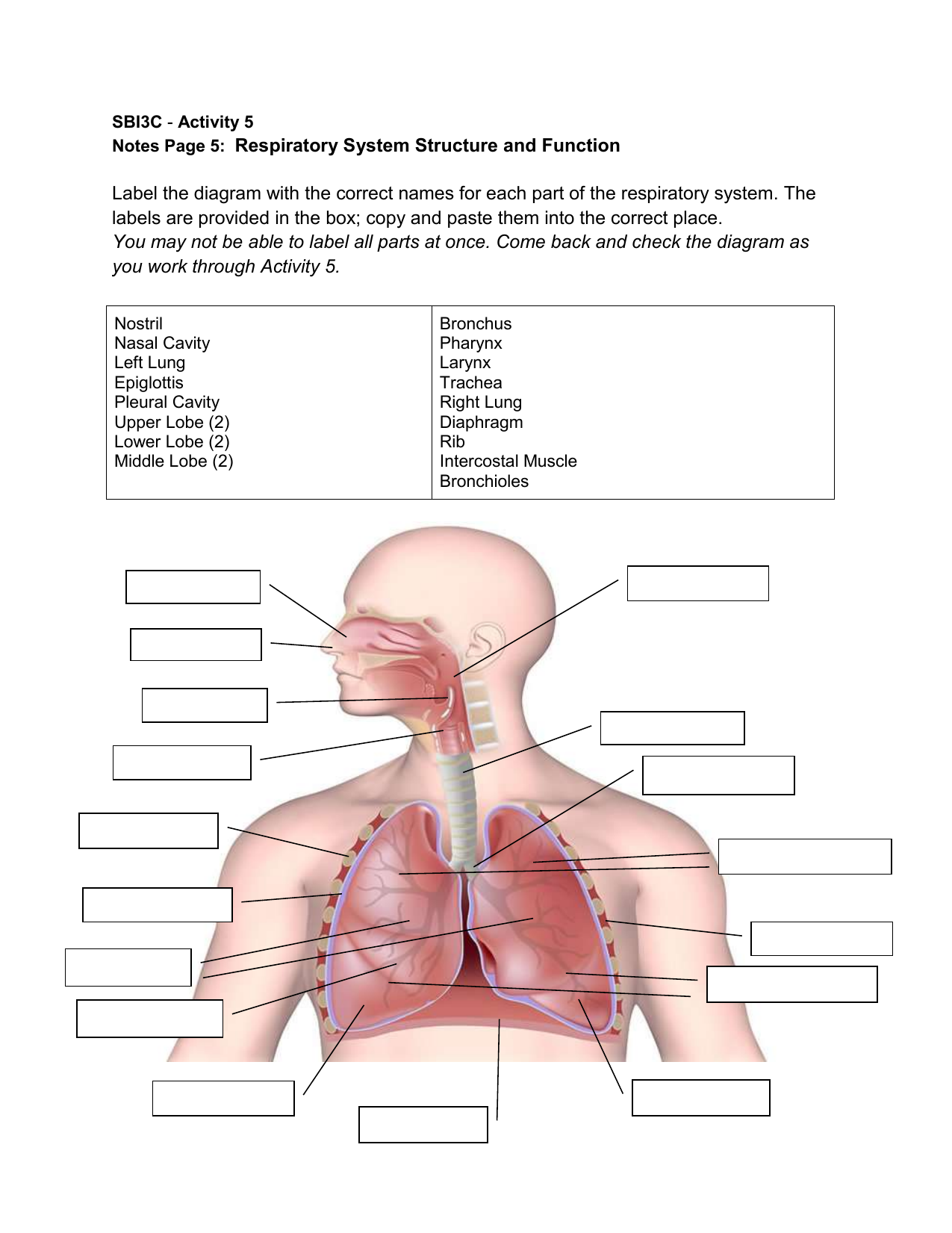
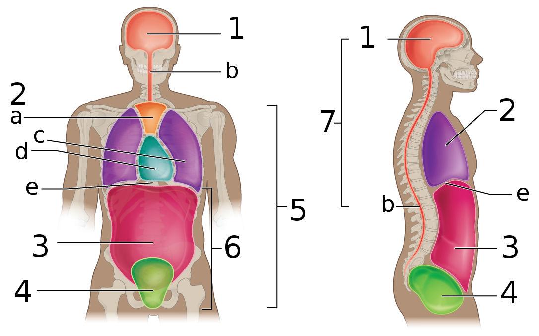
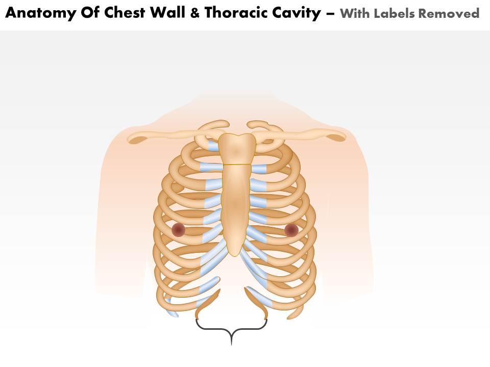
Thoracic cavity | Description, Anatomy, & Physiology | Britannica thoracic cavity, also called chest cavity, the second largest hollow space of the body. It is enclosed by the ribs, the vertebral column, and the sternum, or breastbone, and is separated from the abdominal cavity (the body's largest hollow space) by a muscular and membranous partition, the diaphragm. Anatomy Chapter 1: Labeling Thoracic Cavity Diagram | Quizlet The central portion of the thoracic cavity pericardium The serous membrane surrounding the heart parietal pericardium The aspect of the pericardium that does not touch the surface of the heart visceral pericardium The aspect of the pericardium which covers the exterior surface of the heart pericardial cavity The cavity that surrounds the heart Labeled Diagram of the Human Lungs - Bodytomy Human lungs are located in the thoracic cavity or chest and are enclosed within the rib cage. The two lungs are situated on either sides of the heart and are pinkish in color, especially at a young age. Exposure to the atmosphere and polluted air eventually gives rise to mottled patches, which tint the lungs gray in color. Body Cavities Labeling - The Biology Corner Shows the body cavities from a front view and a lateral view, practice naming the cavity by filling in the boxes. Body Cavities Labeling Name: ______________________________________________ This work is licensed under a Creative Commons Attribution-NonCommercial-ShareAlike 4.0 International License. Answers: Front View: 1. Cranial Cavity 2.
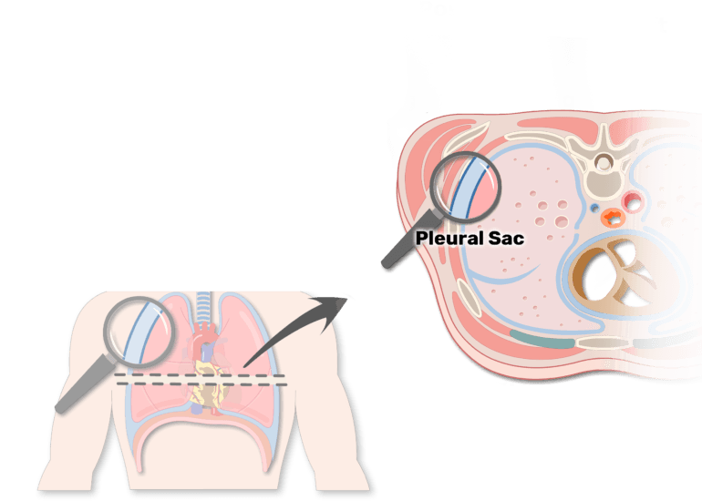
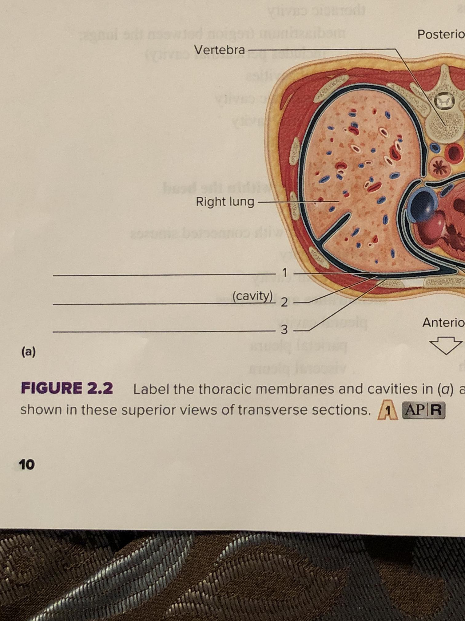
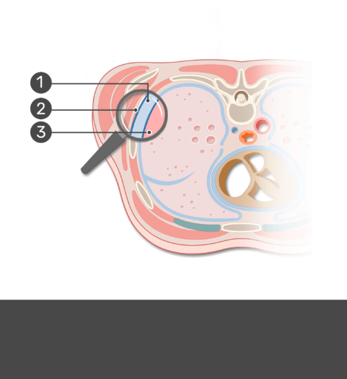
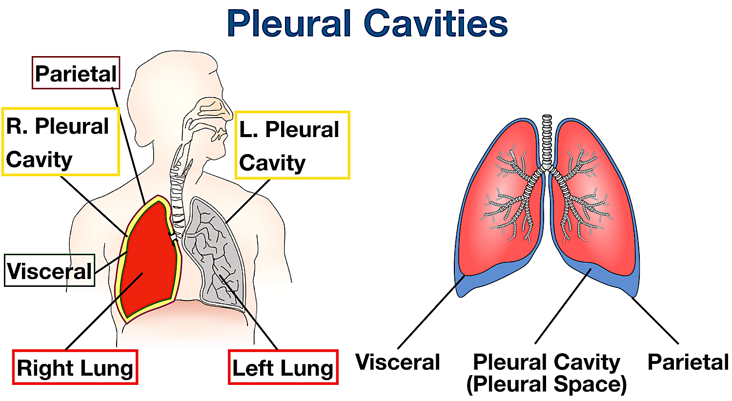
Unit 1 Lab Homework Flashcards | Quizlet Label the regions of the body. Left Down: Cervical Axillary Cubital Antebrachial Crural Right Down: Deltoid Brachial Inguinal Femoral Label the structures of the thoracic cavity. Left Down: Parietal Pleura Pleural Cavity Visceral Pleura Visceral Pericardium Pericardial Cavity Parietal Pericardium Label the directional terms based on the arrows. 1.6 Anatomical Terminology - Anatomy and Physiology 2e - OpenStax The anterior (ventral) cavity has two main subdivisions: the thoracic cavity and the abdominopelvic cavity (see Figure 1.15). The thoracic cavity is the more superior subdivision of the anterior cavity, and it is enclosed by the rib cage. The thoracic cavity contains the lungs and the heart, which is located in the mediastinum. The diaphragm ... Thoracic Cavity - Introduction, Structure, Organs, and FAQs - VEDANTU The thoracic cavity is protected by the thoracic wall. The thoracic wall comprises the rib cage, muscle, and fascia. The mediastinum is known as the central compartment of the thorax cavity. The actual thoracic cavity meaning is that it has two openings that are superior thoracic aperture and lower inferior thoracic aperture. Ch. 19 Circulatory System- heart Flashcards | Quizlet Place the labels in order denoting the flow of blood through the pulmonary circuit beginning with the right atrium and ending in the left atrioventricular valve. The first and last structures are given. Right atrium 1. tricuspid valve 2. right ventricle 3. pulmonary valve 4. pulmonary trunk 5. pulmonary artery 6. lungs 7. pulmonary vein
Thoracic Cage Labeling Quiz - PurposeGames.com This online quiz is called Thoracic Cage Labeling. It was created by member court_48 and has 13 questions. ... Label Lateral View Of The Brain. Science. English. Creator. EllenEllen. Quiz Type. Image Quiz. Value. 10 points. Likes. 17. Played. 56,381 times. Printable Worksheet. Play Now. Add to playlist.
Human Heart - Diagram and Anatomy of the Heart - Innerbody The heart is located in the thoracic cavity medial to the lungs and posterior to the sternum. On its superior end, the base of the heart is attached to the aorta, Continue Scrolling To Read More Below ... The walls and lining of the pericardial cavity are a special membrane known as the pericardium. Pericardium is a type of serous membrane that ...
1.4E: Body Cavities - Medicine LibreTexts The thoracic cavity is the anterior ventral body cavity found within the rib cage in the torso. It houses the primary organs of the cardiovascular and respiratory systems, such as the heart and lungs, but also includes organs from other systems, such as the esophagus and the thymus gland.
Solved Correctly label the following lymphatics of the the | Chegg.com Expert Answer. 100% (12 ratings) From top to bottom First box : Right lymphatic duct. It drains the lymphatic flui …. View the full answer. Transcribed image text: Correctly label the following lymphatics of the the acic cavity. Right subclavian vein Lymphatics of breast Axillary lymph nodes Right lymphatic duct Left lymphatic Thoracic duct duct.
Solved Pre-Lab Exercise 17-2 Label and color the structures - Chegg Anatomy and Physiology questions and answers. Pre-Lab Exercise 17-2 Label and color the structures of the thoracic cavity in Figure 17.1 with the terms from Exercise 17-1 (p. 451). Use your text and Exercise 17-1 in this unit for reference. Label and color the three views of the heart in Figure 17.2 with the terms from Exercise 17-1 (p. 451).
Fetal Pig Dissection - Virtual Anatomy & Diagrams | HST The thoracic cavity is protected by the rib cage and contains the lungs and heart. Use the labeled picture to find the following organs: Lungs - the lungs have multiple lobes and are found on either side of the heart. Heart - the heart is encased in a shiny pericardial membrane; carefully remove this with your scissors or a teasing needle.
Thoracic Cavity - Definition & Organs of Chest Cavity - Biology Dictionary Thoracic Cavity Definition The thoracic cavity, also called the chest cavity, is a cavity of vertebrates bounded by the rib cage on the sides and top, and the diaphragm on the bottom. The chest cavity is bound by the thoracic vertebrae, which connect to the ribs that surround the cavity.
Thoracic cavity - Wikipedia The thoracic cavity (or chest cavity) is the chamber of the body of vertebrates that is protected by the thoracic wall The central compartment of the ...
Solved Award: 0.76 points Label the structures of the - Chegg Award: 0.76 points Label the structures of the thoracic cavity. Parietal pleura Visceral pleura Pleural cavity Parietal pericardium Visceral pericardium Pericardial cavity Reset Zoom This problem has been solved! You'll get a detailed solution from a subject matter expert that helps you learn core concepts. See Answer
Thorax of the dog: normal anatomy | vet-Anatomy - IMAIOS Additional 3D anatomical images at the end are available, showing bones, muscles, vessels, trachea, bronchi and lungs of the thoracic cavity of the dog. 753 anatomical parts have been labeled, separated in different sections: Body parts. Regions. Bones: Vertebral column, Ribs, Sternum, Bones of thoracic limb, Numbering of vertebrae and ribs.
A&P II Flashcards | Quizlet Correctly label the anatomical features of lymphatic capillaries. Correctly label the anatomical features of lymphatic capillaries. Correctly label the lymphatic tissue of the large intestine. Correctly label the following aspects of red bone marrow. Correctly label the following features of the lymphatic system.
Label the structures of the thoracic cavity - Pinterest Award: 0.76 points Label the structures of the thoracic cavity. Parietal pleura Visceral pleura Pleural cavity Parietal pericardium Visceral pericardium ...
Thoracic Cavity - Anatomy | Organs | Functions | 8 Types of Cavities Thoracic cavity meaning in Hindi - वैक्षिक छिद्र What is Thoracic Cavity? The Thoracic cavity (or chest cavity) is that the chamber of the body of vertebrates that are protected by the pectoral wall ( rib cage and associated skin, fascia, and muscle). The central compartment of the thoracic cavity is the mediastinum.
Organs in the Thoracic Cavity - Bodytomy The thoracic cavity is lined by a serous membrane that exudes a thin fluid (serum). The chest membrane, also known as parietal pleura, extends further to cover the lungs. This part of the membrane is known as the visceral pleura. The part of the membrane that covers the heart, esophagus, and the great vessels is known as mediastinal pleura.
Anatomy, Thorax - StatPearls - NCBI Bookshelf - National Center for ... The thoracic cavity contains organs and tissues that function in the respiratory (lungs, bronchi, trachea, pleura), cardiovascular (heart, pericardium, great vessels, lymphatics), nervous (vagus nerve, sympathetic chain, phrenic nerve, recurrent laryngeal nerve), immune (thymus) and digestive (esophagus) systems.
Anatomy of the Thoracic Cavity & Diaphragm - YouTube May 16, 2015 ... This video is about anatomy of the thoracic cavity and diaphragm.University of Sulaimani / College of Medicine.
Body Cavities and Membranes: Labeled Diagram, Definitions - EZmed Body Cavities Labeled Diagram: The dorsal cavity is located in the back of the body (red/stars) and houses the central nervous system including the brain and spinal cord. Ventral Cavity The ventral cavity is the cavity located in the front of the body, which makes sense because ventral means front or anterior.


:watermark(/images/watermark_5000_10percent.png,0,0,0):watermark(/images/logo_url.png,-10,-10,0):format(jpeg)/images/overview_image/38/468rAQ00megI9kRFqvOT1A_lungs-in-situ_english.jpg)












Komentar
Posting Komentar